0452 2536 576
+91 9655 015 015
info@aasastha.com

-
-
No: 14, Gokhale Road
Opp Booma Hospital Chokkikulam, Madurai.
Contact Info
-
Address
No: 14, Gokhale Road
Opp Booma Hospital Chokkikulam, Madurai. -
Phone
0452 2536 576
+91 9655 015 015
info@aasastha.com -
Email
info@aasastha.com
© AASASTHA 2022, All Rights Reserved.
Digital Imaging Basics
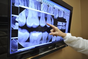
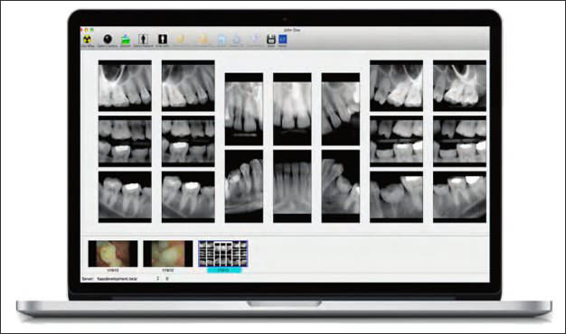
The production of digital images requires a process called Analog to Digital Conversion (ADC). Digital imaging is a radiographic technique that utilizes a wired or wireless hard sensor or phosphor plate sensors known as a receptor, instead of film. Digital images consist of pixels organized in a matrix of rows and columns. ADC consists of two steps: sampling and quantization. Sampling is a small range of voltage values that are grouped together as a single value. Once sampled, every sampled signal is assigned a value. For the dental provider to see the digital images, the computer must organize the pixels in their correct locations and display shades of gray that corresponds to the number that was assigned. This step is known as quantization.5
In digital imaging, the x-ray machine is still used, but the image is converted from analog to digital. When the photons from the x-ray tubehead strike the sensor, the analog image is converted to a digital image and then transferred to the digital imaging software (Figure 1). The digital images are composed of pixels or rows and columns of images aligned to represent the intensity (gray level) of the image. There is a value assigned to each pixel that represents the intensity or gray level of each location in the image.9 The computer software organizes the pixels and displays the dentistry of the object via multiple shades of gray. Due to pixel technology in digital sensors, anatomy such as bone trabeculation, lamina dura, and DEJ are easier to diagnose due to high contrast in digital imaging.
Digital Imaging in Dentistry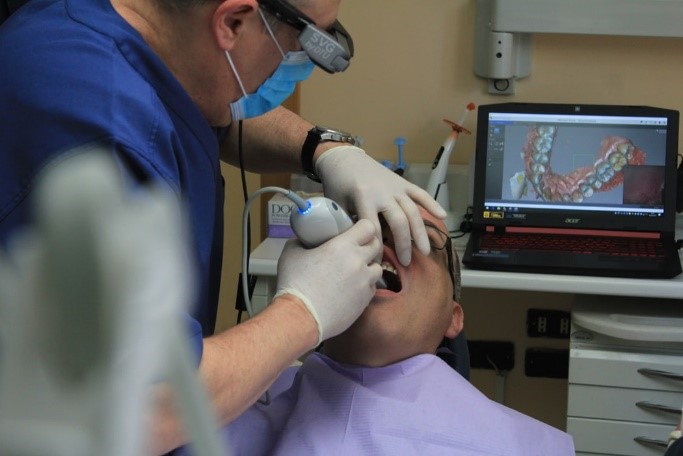
Digital Imaging Radiation Exposure
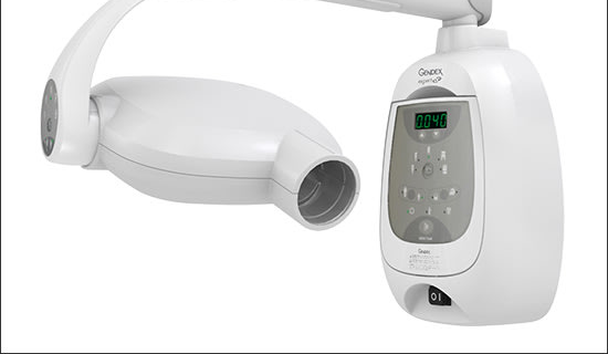
Even though digital imaging requires less radiation than radiographic film, radiation is still produced at the source, e.g., dental x-ray machine. Digital imaging reduces the amount of radiation to the patient due to the sensitivity of the digital imaging sensors. Depending on the radiographic film used in the dental practice, e.g., D- or F-speed, radiation exposure time can be reduced as much as 90% when changing from radiographic film to digital sensors.1,5,6,7 For example, an exposure time required to produce a digital image is 0.05 seconds, where a radiographic film would require an exposure time of 0.2 seconds. Standard radiation safety procedures still requires the dental provider be at least 6 feet from the x-ray tubehead and the patient be covered in a lead apron with a thyroid collar.


Meet Our Specialist

Dr. A. Bama Selvakumar., MDS
Orthodontist and Dentofacial Orthopedician
Dr.R.P.Gomathi MDS
Periodontist & Implantologist